 How to identify a mineral ? - Part 2
Introduction :
As seen previously, although the identification criteria are numerous, it is not always easy to identify a mineral, especially when it is small. Here we will deal with the main modern identification methods.
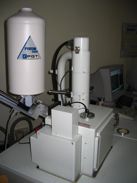
The scanning electron microscope (SEM) :

It is undoubtedly the favorite tool of micromineral collectors, it is a device that is capable of producing high resolution images of the surface of a sample using an electron source. An electron gun bombards the sample which in response re-emits particles that can be analyzed by different detectors :
Secondary electron detector (GSE) :
It makes it possible to have a black and white image of the surface of the sample. This makes it possible to appreciate the morphology of crystals, even very small ! Opposite an example of an image of crystals impossible to identify with a binocular magnifying microscope because they are too small.
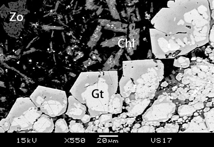
Backscattered electron detectors :
The quantity of electrons captured by the atoms encountered and therefore the quantity of backscattered electrons which emerge from the sample depends on the chemical nature of the layers of material crossed. The electron emission rate increases with the atomic number. A chemical contrast is therefore obtained, the zones containing light atoms appear darker than the zones which contain heavy elements. It should be noted that to be really interesting this technique is used most of the time on polished sections of rock or minerals.
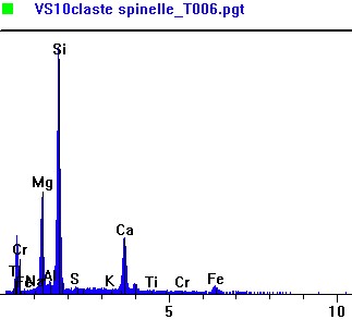
Elemental analysis by X-ray spectrometry (EDS detector) :
The energy of the X-rays emitted during the de-excitation of the atoms bombarded by the electron gun depends on their chemical nature. By analyzing the X-ray spectrum, we can have an elementary analysis, that is, know what types of atoms are present in the sample and in what quantity. The beam scanning the sample, we can even draw up a chemical map, with however a resolution much lower than the image in secondary electrons.
The SEM allows one-off identification to be carried out efficiently and quickly. Samples can be analyzed raw, without prior preparation. It is possible to study large samples larger than 20 cm. The device with all the detectors mentioned above costs a little over 300,000€, an hour of assisted analysis is billed between 50€ and 80€. In recent years, we have seen the appearance of so-called "tabletop" SEMs at lower prices (around 50,000€), they are more limited than conventional SEMs, their sample locks are much smaller, but it is nonetheless not less interesting for all that !
On the other hand, the SEM only allows the analysis of the major elements of a sample in a non-destructive manner. The results obtained are not considered "precise" enough to be published in scientific journals.
The electronic microprobe :
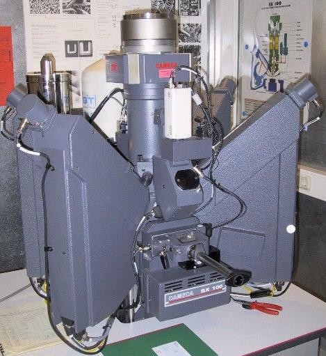
It consists in bombarding a sample with electrons and in analyzing the spectrum of X-rays emitted by the sample under this stress. X-rays are diffracted on a crystal and analyzed by a WDS detector (different from the EDS of the SEM).
Depending on the number of diffraction crystals equipped on the machine it can analyze up to 4 chemical elements at the same time. This requires knowing which chemical elements to analyze and juggling diffraction crystals, since a crystal does not allow the analysis of all the chemical elements in the periodic table. It is therefore essential to have already analyzed the specimens with the SEM. The samples analyzed must be polished and metallized (coated with a thin layer of carbon using a metallizer). One can wonder what is the advantage of this machine compared to the SEM, well it is the precision ! The microprobe allows precise punctual quantitative analyzes of major elements but also of trace elements (contained in very small quantities in the mineral) of very small objects. The results are presented in the form of tables of figures.
The results of microprobe analyzes are precise and publishable. The device costs around 700,000 €. Analyzes are long to set up and expensive. They require a good knowledge of the sample and are partially destructive in the sense that one has to prepare the sample.
X-ray diffraction (XRD) :

The principle is to bombard a sample with X-rays which are deflected by the crystal structure of the mineral.
The sample is prepared as a flattened powder in a cup, or as a solid, flat wafer. X-rays are sent to this sample, and a detector goes around the sample to measure the intensity of the X-rays in the direction. For practical reasons, the sample is rotated at the same time, or possibly the tube producing the x-rays is rotated.
Modern devices identify the mineral or minerals if it is a mixture from a database. The spectrum costs 30€.
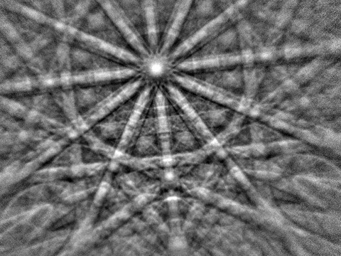
The transmission electron microscope (TEM) :
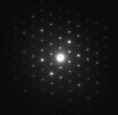
It works on the same principle as the SEM except that the electron beam is "transmitted" through a very thin sample. This device allows fine and precise punctual analyzes but above all to have punctual information on the crystalline structure of the mineral ! Opposite a diffraction image of a phyllosilicates of a pseudotachylite vein.
It requires an heavy preparation but also the destruction of the samples. Either the mineral must be powdered or it must undergo ionic thinning.
Raman :
Raman spectroscopy is non-destructive, it allows to characterize minerals and even better : their inclusions !
A beam of monochromatic light (laser) is sent on the sample to be studied and the scattered light is analyzed after having been collected by another lens and sent into a monochromator allowing its intensity to be measured using a detector.
Raman or micro-Raman microspectroscopy is a microscopic measurement technique : by focusing the laser beam on a small part of the environment, the properties of this environment can be probed over a volume of a few µm3.
Identifications by this means are carried out by comparison of spectra of known minerals.
Conclusion :
These different modern analysis methods now make it possible to provide certainty when it comes to identifying crystals. And it is also what allows collectors of micro-minerals to give precise names to their discoveries.
Références :
- Wikipédia